Cell Culture 101: Tips for Successful Cell Culture
February 09, 2017, at 12:00 PM ETAbstract
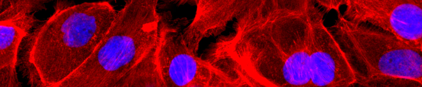
ATCC is widely recognized as the expert in cell culture and production, having the largest supply of cryopreserved cells in the world. In this webinar, we will provide best practices for culturing of cells, from continuous cell lines to primary cells. The information delivered will cover all aspects of successful culture initiation, expansion, authentication, and cryopreservation.
Key Points
- ATCC is world renowned as the definitive expert on all aspects of cell culture and production
- Using misidentified or cross-contaminated cell lines in experiments can invalidate research efforts, therefore authenticating cell lines should be part of your cell culture work flow
- When culturing specialty cells, such as stem cells or primary cells, certain considerations regarding the choice of media and reagents must be taken
Presenters
Kevin Grady, BS
Manager, Product Management, ATCC
Kevin Grady is the Manager of Product Management at ATCC. He has been with ATCC for 8 years; prior to ATCC, he held positions at Lonza as Global Product Manager and Director of Scientific Support. Kevin has a long history in the life science industry additionally serving as Director of Scientific Support at Amaxa and Manager of Technical Support at Life Technologies. Mr. Grady has always found great satisfaction in helping researchers learn how to use available products and tools to be more productive and successful in reaching their research goals.
Steven Budd, MS, MBA
Product Specialist, ATCC
Steven Budd is a Product Specialist that manages the cell culture reagents at ATCC. He has 6 years of experience in the product management of scientific tools. Before that, he gained 4 years of experience in biomedical research and cell culture as a research specialist at the University of North Carolina at Chapel Hill. Mr. Budd has a M.S. in Biology from the University of North Carolina at Wilmington and an M.B.A. from North Carolina State University.
Questions and Answers
Can we use contaminated culture after treating with antibiotics?
We do not recommend using cells after they have been contaminated. In addition to being difficult to get rid of the contaminant, cells may also become physiologically compromised from exposure to the contaminant. If a contamination does occur, these cultures should be discarded, and new cultures should be grown from master stocks that are verified to be contamination-free.
If antibiotics and antimycotics are not recommended, this means that we must rely heavily on aseptic techniques and good practice to prevent contamination. If there is any contamination, do we have to throw away all the cells and start over from the master stock?
Best practices in cell culture include using strict aseptic techniques when handling cells. ATCC does not use antibiotics or antimycotics for routine cell culture. Long-term use of antibiotics or antimycotics may mask the presence of low levels of microbial or mycoplasma contamination. In addition, some antibiotics and antimycotics are toxic and may affect the recovery and proliferation of some cell lines.
However, one may elect to introduce antibiotics for short periods to primary cultures or as a safeguard while propagating specific valuable stocks to produce working stocks. If you do elect to use an antibiotic in your medium, ATCC recommends using a Penicillin-Streptomycin solution at a final concentration of 50-100 I.U./ml penicillin and 50-100 µg/ml streptomycin. ATCC offers a Penicillin-Streptomycin solution (ATCC 30-2300); this sterile solution can be added at 0.5 to 1 ml of solution per 100 mL of cell culture media for a final concentration of 50 to 100 I.U./mL penicillin and 50 to 100 µg/mL streptomycin.
If a contamination does occur, new cultures should be grown from master stocks that are verified to be contamination-free. We do not recommend using cells after they have been contaminated. In addition to being difficult to get rid of the contaminant, the cells may also become physiologically compromised from exposure to the contaminant.
Do you have a recommended animal serum-free alternative for FBS in media?
ATCC does not supply serum alternatives or specialty serum-free media. Other suppliers may provide serum-free media for specific applications or cell types. A good all-purpose, serum-free medium is AIM-V Medium from Thermo Fisher Scientific. As for serum substitutes, you can add an ITS Solution (insulin, transferrin, and selenium) to your basal medium. Alternatively, you can try Human Platelet-derived Lysate. Used separately or in conjunction, this could allow for a slow transition to serum-free conditions or, at least, reduced-serum conditions.
What additional considerations could you suggest for the culture of THP-1 cells (ATCC TIB-202) and for avoiding contamination?
The best way to avoid contamination is to use good aseptic technique. We suggest that you use mycoplasma testing and STR analysis to ensure that your culture is free of contamination, or start with a new vial from ATCC (THP-1, ATCC TIB-202). When handling the cell line, initiate the culture and expand it for 3 to 4 passages; freeze down your own stock at passage 3 or 4. You can grow the culture for about 20 passages, checking for mycoplasma every 4-6 weeks. After 20 passages, you should start up a new culture from your frozen stock.
THP-1 cell line cultures are seeded at 2-4 x 10⁵ viable cells/mL. Subculture when the cell concentration reaches 8 x 10⁵ cells/mL. Do not allow the cell concentration to exceed 1 x 10⁶ cells/mL. The base medium for this cell line is RPMI-1640 Medium (ATCC 30-2001). To make the complete growth medium, add the following components to the base medium: 2-mercaptoethanol to a final concentration of 0.05 mM and Fetal Bovine Serum (FBS; ATCC 30-2020) to a final concentration of 10%.
What can cause adherent cells to detach from the plate?
Most often, this is due to suboptimal medium pH and incorrect buffering caused by an imbalance in the incubator carbon dioxide percentage and the amount of sodium bicarbonate present in the basal media. Most ATCC-formulated basal medium contains 1.5 g/L bicarbonate, for which 5% CO2 is recommended. When using an alternative DMEM formulation that contains 3.7 g/L bicarbonate, the cells should be incubated in 10% CO2; this increase is necessary to ensure that the medium is buffered properly.
In addition, cell detachment can also be caused by the confluency/density of cells in the flask; if there are too many cells, they can start lifting off due to lack of substrate.
Finally, the concentration and time of exposure of the dissociation enzymes used on cells during subculture, incomplete inactivation or removal of dissociating enzymes, and cell clumping can also delay or prevent cell attachment.
What is the best way to change media for spheroid culture models?
Half of the media should be pipetted off and replaced with a one-half volume of fresh media; the spheroid should be left undisturbed at the bottom of the well. There are also special 96-well plates that have a ledge within the well specifically for this purpose, so you may want to start your culture in one of these types of plates.
What is the definition of the working passage number? What are your recommendations for passage number for specific cells?
A passage number simply refers to the number of times the cells in the culture have been removed from one vessel into another (subcultured). The working passage number varies between cell types: a continuous line can be in culture for up to 20 passages, a primary cell for up to 8-12 passages, and a stem cell for up to 30 passages.
What types of experiments can I use to troubleshoot large variations in transient transfection expression between different cell passage numbers?
For starters, you want rule out contamination issues that may be present in the later passage material as contamination can suppress transfection efficiency; “younger” material (lower passage number) will generally exhibit higher transfection efficiency. Depending on the cell type, a >3 passage difference can have a great effect (a greater effect is likely in primary cells and iPSCs).
To check for passage differences, set up an experiment using cells at low, medium, and high passage number (the more samples you have the better time course you can run); complex a small, easily expressed reporter plasmid, like GFP or beta-gal, with your transfection reagent; and split the complexes up for transfection of the two samples. Wait 24 hours and run your analysis. Review your results and establish a passage number window appropriate for the transfection of your cell line/type.
When preparing freeze media, what concentration of DMSO do you recommend?
In general, we use 10%, but we have heard that potentially dropping it to 7.5% might be better. Concentrations of 7.5% or 10% DMS are often used when freezing cells; ATCC routinely recommends 10% DMSO. Cells should be handled quickly during this process to limit the exposure time to DMSO.
Why is it recommended to harvest/subculture cells at 80% confluence and not wait until the culture reaches 100 % confluence?
Normal cells should be subcultured as soon as confluence is reached, or prior to reaching confluence. At 100% confluence, cells begin to enter a “plateau” phase of growth and stop growing and start to die. Harvesting or subculturing the cells at 80% confluence, or when they are still in the exponential (log) phase of growth, will yield the maximum number of healthy, viable cells.
Will freezing the cells directly in -80°C freezer cause damage to the cells? Is it better to reduce the temperature by -20°C and then move to the -80°C?
As the cell suspension is cooled below the freezing point, ice crystals form and the concentration of the solutes in the suspension increases. Intracellular ice can be minimized if water within the cell is allowed to escape by osmosis during the cooling process. A slow cooling rate, generally -1°C per minute, facilitates this process. However, as the cells lose water, they shrink in size and quickly lose viability if they go beyond a minimum volume. The addition of cryoprotective agents such as glycerol or dimethylsufloxide (DMSO; ATCC 4-X) will mitigate these effects. The standard procedure for cryopreservation is to freeze cells slowly in a medium that includes a cryoprotectant until they reach a temperature below -70°C. Then, transfer the vials to a liquid-nitrogen freezer to maintain them at temperatures below -130°C. You can use a commercially available freezing container, such as the CoolCell LX Alcohol-free Cryopreservation Container (ATCC ACS-6000). These containers are specifically designed to cool at -1°C/minute when placed directly into a -70°C cooler or freezer.