An Authenticated In Vitro Model for Prostate Microenvironment Studies That Utilizes Prostate Epithelial Cells and Stromal-derived Cells
AACR Annual Meeting 2018
Chicago, Illinois, United States
April 14, 2018Abstract
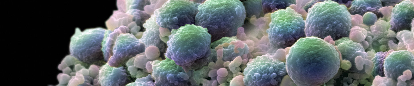
Prostate cancer remains one of the most common cancers diagnosed in men and one of the leading causes of cancer death in men. Tumor development and progression have been shown to be highly influenced not simply by the genetic makeup of a cell, but by its surrounding stroma (particularly fibroblasts). It has been demonstrated that prostate cancer–associated fibroblasts (CAFs, which are located marginal to the prostate tumor) differ from prostate normal–associated fibroblast (NAFs, which are located distal to the prostate tumor) with regard to their contribution to tumor progression. However, human prostate cancer in vitro model systems have focused largely on prostate cancer epithelial cells exclusively. A need exists for a more physiologically relevant human cell model system to study prostate cancer progression within the context of its tumor microenvironment.
In this study, we utilized prostate CAFs, prostate NAFs, and normal prostate epithelial (PrE) cells; all 3 lines were immortalized by human telomerase reverse transcriptase (hTERT) alone and then were cryopreserved, thawed, and continuously passaged without any indication of a decrease in growth rate. All cell lines expressed appropriate specific cell lineage markers for either fibroblasts or epithelial cells. Fibroblasts expressed TE7 and alpha smooth muscle actin (a-SMA), while PrE cells expressed cytokeratin 18, low levels of prostate specific antigen (PSA), and high levels of P63 throughout their passage; all characteristics were in accord with their primary cell counterparts. Next, cell proliferation was measured under the influence of CAFs and NAFs for various prostate-derived epithelial cells. The effects of stromal cells on prostate cell proliferation was cell line–dependent. CAF cells promoted the growth of PrE, DU145, and the unrelated epithelial cell A549, while NAF cells inhibited their growth. Meanwhile, CAF cells inhibited the growth of the viral transduced RWPE1 cell line, while NAF cells promoted the growth of the cancer-derived prostate epithelial cell LNCap. This study demonstrates that these 3 hTERT-immortalized cells from human prostate are a valuable model system for the study of prostate cancer cell progression and tumor microenvironment studies.
Download the poster to explore the use of hTERT-immortalized cells as models in prostate cancer research.
Download