TIME cells maintain their normal growth rate and endothelial character at late passage when grown in ATCC’s VCBM-VEGF medium, but not in the previously recommended Lonza EGM-2 MV BulletKit medium.
Introduction
The telomerase-immortalized microvascular endothelial (TIME) cell line was generated by hTERT-immortalization of primary neonatal foreskin microvascular endothelial cells. These cells exhibit a normal karyotype, extended lifespan in culture, and endothelial characteristics that make them an ideal model for investigating many aspects of endothelial cell biology. However, their growth rate slows and they lose their endothelial character at later passages when cultured in the recommended Lonza EGM-2-MV BulletKit medium. In contrast, their growth rate and endothelial character are maintained at late passage when grown in ATCC Vascular Cell Basal Medium plus VEGF (VCBM-VEGF). Thus, ATCC now recommends VCBM-VEGF as the preferred medium for growing TIME cells.
Download a PDF of this application note
Download NowMaterials and methods
TIME Cells (ATCC CRL-4025) at passage 2 (P2) or passage 12 (P12) were seeded at 5 × 10³ cells/cm2 in 6-well plates containing either ATCC Vascular Cell Basal Medium (VCBM-VEGF) (ATCC PCS-100-030) plus Endothelial Cell Growth Kit-VEGF (ATCC PCS-100-041) or Lonza EGM-2MV BulletKit (CC-3202, Lonza, Walkersville, MD). Growth rate was assessed as population doubling levels (PDL) or as percent confluence over time monitored using an IncuCyte FLR (Essen BioScience, Ann Arbor, MI)]. CD31 expression was examined by fluorescence microscopy of TIME cells stained using an α-CD31 monoclonal antibody (R&D Systems, Minneapolis, MN) at 10 μg/mL overnight, and Alexa Fluor 594 Goat Anti-Mouse IgG (H+L; Life Technologies, Grand Island, NY).
Results and discussion
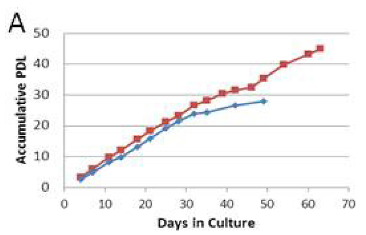
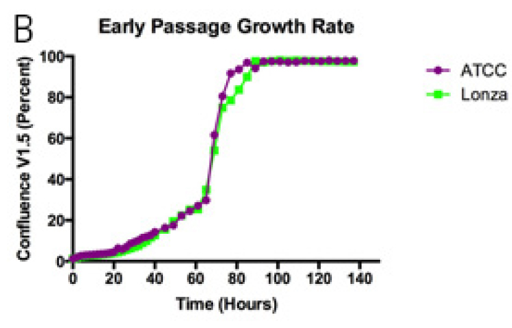
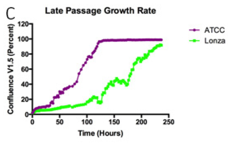
In ATCC medium, TIME cells reached a PDL of ≥45 while in the Lonza medium they only reached a PDL of 25 before their growth rate slowed (Panel A). To examine this further, we monitored the growth rate of TIME cells at early (P2) and late passage (P12) in the different media formulations. P2 TIME cells cultured in either medium reached 100% confluency by day 4 in culture (Panel B). At P12, the growth rate of TIME cells grown in the ATCC medium was similar to the P2 cells, taking about 5 days to reach confluence (Panel C, red curve). In contrast, P12 cells grown in the Lonza medium took nearly 10 days to become fully confluent (Panel C, blue curve), indicating a substantial reduction in their rate of growth.
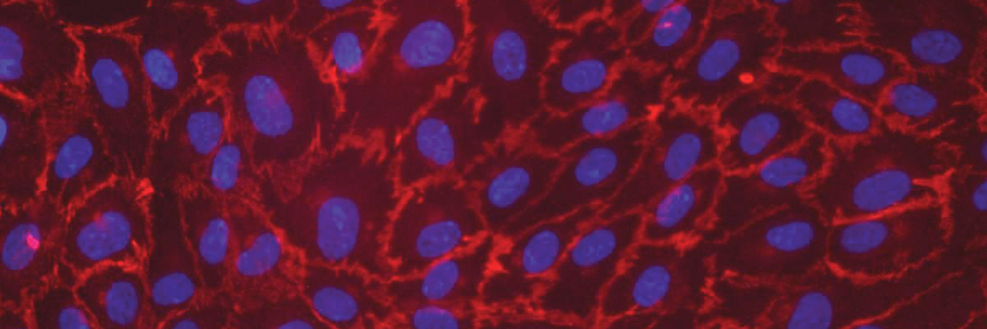
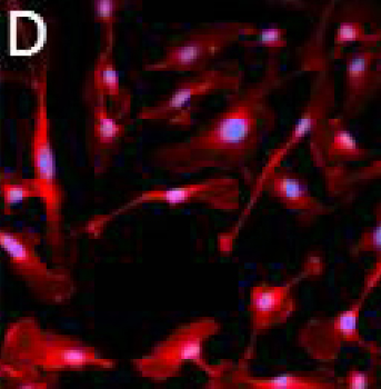
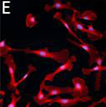
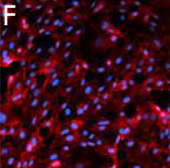
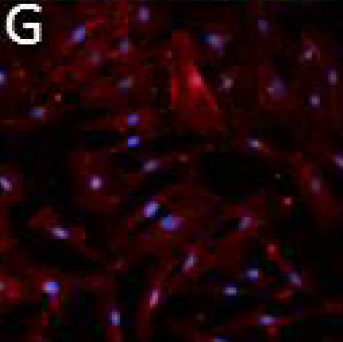
To determine whether TIME cells retained their endothelial characteristics at late passage, cells were stained for the endothelial cell marker PECAM/CD31. At P2, TIME cells grown in either medium showed robust expression of PECAM/CD31 (Panels D, E, respectively), and at P12 TIME cells in ATCC medium maintain a high level of PECAM/CD31 expression (Panel F). In contrast, PECAM/CD31 expression is markedly reduced in P12 TIME cells grown in the Lonza medium (Panel G), suggesting that P12 TIME cells begin to lose their endothelial character after extended culture in the Lonza medium.
 |
 |
 |
 |
 |
 |
 |
Conclusion
TIME cells cultured in ATCC VCBM-VEGF medium retain their robust growth and endothelial characteristics and can be cultured longer than in the previously recommended medium. Therefore, ATCC now recommends ATCC VCBM-VEGF medium as the preferred medium for the growth of TIME cells.
Download a PDF of this application note
Download Now