The Problem
Mycoplasmas are frequent contaminants of cell cultures and bioprocessing fluids. It is well established that mycoplasma contamination of continuous cell cultures ranges from 15-35%, with primary cell cultures exhibiting a minimum 1% contamination rate. Mycoplasma contamination can be caused by poor culturing practices or malfunctioning laboratory equipment. As an example of poor culture practice, reusing pipet tips may transfer mycoplasma-infected media into otherwise sterile media, propagating the spread of the contaminant. On the other hand, faulty laminar flow could disperse mycoplasma-containing dust and aerosols throughout a biosafety cabinet, thereby contaminating all of the media and cells within.
There are over 190 species of mycoplasma, but only 20 distinct species of human, bovine and porcine origin have been identified in cell culture. Of those twenty, eight species account for approximately 95% of all mycoplasma contamination in cell culture, including M. arginini (bovine), M. fermentans (human), M. hominis (human), M. hyorhinis (porcine), M. orale (human), M. pirum (human), M. salivarium (human), and Acholeplasma laidlawii (bovine).
Mycoplasma can affect the phenotypic and functional characteristics of cells in vitro, including:
- Chromosomal aberrations
- Disruption of nucleic acid synthesis
- Changes in membrane antigenicity
- Inhibition of cell proliferation and metabolism
- Decreased transfection rates
- Changes in gene expression profiles
- Affects virus production
- Cell death
Best Practices to Protect Your Cells
While mycoplasma contamination is difficult to treat, it can be prevented by implementing a few best practices. For example, using proper aseptic techniques and practices such as wearing personal protective equipment (PPE) and working in a certified vertical laminar flow hood will diminish the chances of contamination from the environment. In addition, quarantining new cell lines of any origin reduces the risk of cross contamination. Employing standard antibiotics does not protect cell cultures against mycoplasma contamination: Penicillin has no effect on mycoplasma since mycoplasma lack a cell wall; streptomycin inhibits about half the mycoplasma strains but is ineffective against many others; gentamycin is generally ineffective at the concentrations routinely used in cell culture. In fact, mycoplasmas are generally resistant to most antibiotic mixtures commonly used in cell culture. Because many antibiotics have no effect on mycoplasma, don’t indiscriminately use antibiotics to prevent contamination. While there is no chemical preventative, implementing good cell banking practices will allow you to quickly recover if your working cell stocks becomes contaminated. Adhering to the seed stock principle ensures that you always have fresh stocks of cells on hand to replenish a contaminated working cell bank. In addition, confirming that all of the media, sera, and reagents you use in your experiments are obtained from mycoplasma-free sources will safeguard your cells from contamination by external sources. Finally, routinely testing your cells for mycoplasma contamination will ensure that you can trust your experimental results.
Early Detection Leads to Prevention
The best protection against mycoplasma contamination is to quickly identify contaminated cultures and reagents through routine testing. Contamination of cell substrates used in the production of biopharmaceuticals poses a potential safety risk for patients and presents a serious economic risk for manufacturers in the event of batch adulteration or a product recall. To minimize these risks, routine testing for mycoplasma is performed throughout the product manufacturing and development process. These testing methods include direct culture, indirect culture, and PCR-based detection.
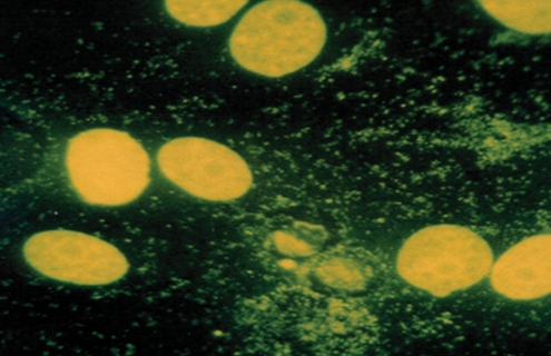
In the direct culture method, an agar plate is inoculated with the culture in question and incubated for a period of four to five weeks. The plates are then monitored for the appearance of fried egg-shaped colonies, which signals a positive result. By contrast, the indirect culture method simply requires culturing the cells under normal growth conditions. Once the cells reach an appropriate confluence, they are stained with Hoechst 33258, a fluorescent dye that binds specifically to DNA. The appearance of filamentous staining in the cytoplasm of the cells in question indicates a positive finding.
For more rapid and qualitative testing, a PCR-based method exists that can quickly and reliably identify over 60 species of Mycoplasma, Acholeplasma, Spiroplasma, and Ureaplasma, including the top eight species most likely to affect cell cultures: M. arginini, M. fermentans, M. hominis, M. hyorhinis, M. orale, M. pirum, M. salivarium, and A. laidlawii. ATCC offers this method as either a mycoplasma testing service or a kit. Here, our method uses universal PCR primers targeted to the 16S rRNA gene, a touchdown PCR protocol to increase sensitivity, and a unique thermostable DNA polymerase to increase specificity. Importantly, the PCR-based method of mycoplasma detection meets the standards of the European Pharmacopeia 2.6.7 guidelines (Biologicals 38, March 2010).
Cell Line Contamination – Mycoplasma Prevention and Detection
Mycoplasma contamination significantly affects cell line physiology, resulting in erroneous data and misinterpretation of results. Watch our on-demand webinar to explore the best practices for routine testing for mycoplasma contamination.
Watch the Webinar
Don't Risk Your Data
Misidentified and contaminated cell lines undermine your experimental results and discredit preclinical studies. Your research is too important to risk. Be a part of the movement to raise credibility in science and order your cell line authentication and mycoplasma testing services today.
Check your cellsMycoplasma quality control of cell substrates and biopharmaceuticals
Mycoplasma contamination constitutes a serious concern for cell culturists as these bacterial strains are a common cause of cell line contamination affecting roughly 15-35% of cell cultures and endangering almost all aspects of cell physiology.
Learn More