The transition from an epithelial to mesenchymal cell phenotype (EMT) and its reverse mesenchymal-epithelial transition (MET) are essential for wound healing and many other normal, physiological, developmental processes; these fundamental cellular changes have also been implicated in cancer metastasis and organ fibrosis. During the EMT process, epithelial cells lose their apical-basal polarity and cell-cell adhesive properties and gain migratory and invasive properties, thereby exhibiting the characteristics of mesenchymal stem cells. In contrast, during the MET process mesenchymal cells lose their motility and show an increased number of cell-cell adhesions.
Scientists have implicated EMT in metastasis and are thus targeting EMT/MET pathways as a potential strategy for cancer treatment. As researchers investigate the physiological mechanisms implicated in metastasis, they need access to credible cell models that demonstrate the forward and retrograde transition from an epithelial to a mesenchymal phenotype. To help researchers in the fight against cancer and the development of new treatment options, ATCC scientists created reporter cell lines that enable the real-time monitoring of EMT.
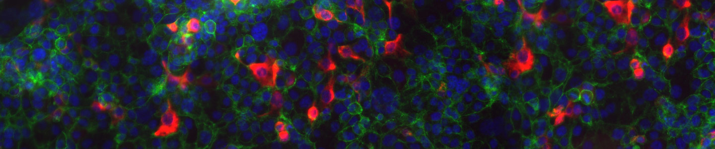
The HCT116 VIM RFP, A549 VIM RFP, and MDA-MB-231 VIM RFP cell lines were generated by using CRISPR/Cas9 genome-editing technology to integrate a red fluorescent protein (RFP) reporter molecule before the stop codon at the last exon of the endogenous vimentin gene. This genetic manipulation enables researchers to track the EMT status of cells by monitoring RFP expression and to gain insight into this phenomenon in colon, lung, and breast cancer models.
EMT Research Solutions
Reporter-labeled Cell Lines
Epithelial-mesenchymal transition reporter cell lines not only provide a model for understanding metastasis, they also deliver a robust platform for developing new cancer therapies.
Learn More
Discover Models for Breast Cancer Metastasis
Despite evidence linking EMT with cancer metastasis, EMT has not been an active target for therapeutic development. This is due in part to the lack of appropriate in vitro models. Watch our presentation to explore the development of a metastatic breast cancer reporter cell line model.
See the PresentationVIM-RFP Reporter Line for Colorectal Cancer
Evidence indicates that EMT displays intermediate states in response to signaling pathway molecules and microRNAs (mRNAs). There is also a body of research indicating that EMT plays an important role in cancer cell dissemination and metastasis and therefore, targeting EMT is considered a novel opportunity in anticancer treatment and drug development.
Explore the Research